
Normal anatomy of the brain on CT and MRI with a few normal variants Practical Neurology
The cerebellum (infratentorial or back of brain) is located at the back of the head. Its function is to coordinate voluntary muscle movements and to maintain posture, balance, and equilibrium. More specifically, other parts of the brain include the following: Pons.

Brain and face CT interactive anatomy atlas eAnatomy
IMAIOS and selected third parties, use cookies or similar technologies, in particular for audience measurement. Cookies allow us to analyze and store information such as the characteristics of your device as well as certain personal data (e.g., IP addresses, navigation, usage or geolocation data, unique identifiers).

Brain and face CT interactive anatomy atlas eAnatomy
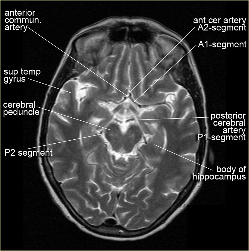
Different areas of the brain are supplied by the anterior, middle and posterior cerebral arteries in a predictable distribution. The posterior fossa structures are supplied by the vertebrobasilar arteries.. The arteries of the brain are not well visualised on conventional CT, but a knowledge of the areas of the brain they supply is helpful in determining the source of a vascular insult.
The Radiology Assistant Brain Anatomy
Key points Grey matter appears grey White matter appears blacker The brain consists of grey and white matter structures which are differentiated on CT by differences in density. White matter has a high content of myelinated axons. Grey matter contains relatively few axons and a higher number of cell bodies.

Normal anatomy of the brain on CT and MRI with a few normal variants Practical Neurology
Last updated: 29 June 2022. Basic radiological anatomy of the brain and spine with annotated CT and MRI images covering the brain, including the brainstem structures and ventricles, and whole spine.
MRI Brain Anatomy
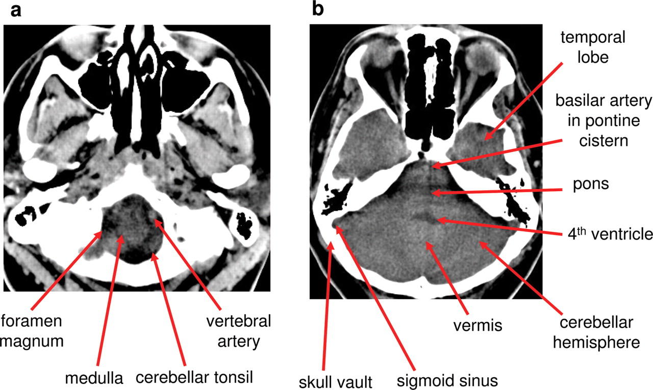
Normal CT head (with labels) Annotated image The labeled structures are (excluding the correct side): foramen magnum medulla oblongata vertebral artery cerebellar tonsil premedullary cistern internal jugular vein basilar artery sigmoid sinus petrous internal carotid artery in the carotid canal cerebellar hemisphere external auditory canal
The Radiology Assistant Brain Anatomy
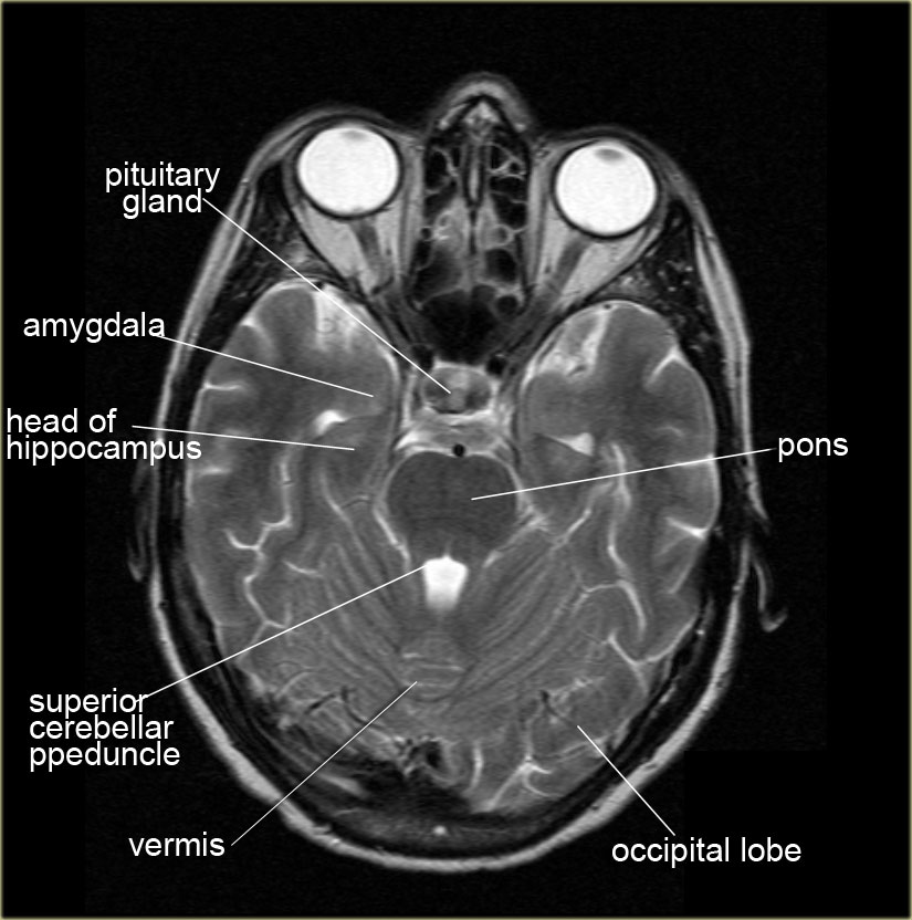
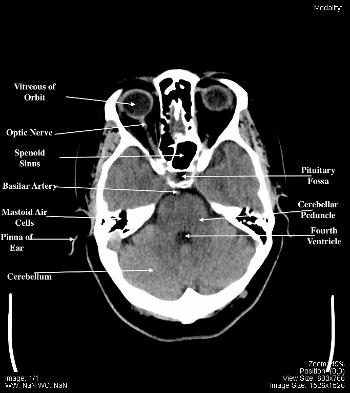
Identifying the pituitary gland on brain CT. In adults over 40 years of age, the pituitary gland sits inside the sella turcica and does not normally extend above the dorsum sellae (the posterior wall of the sella turcica). On a normal brain CT scan, you will see a fluid space in the region just anterior to the dosum sellae.

Normal anatomy of the brain on CT and MRI with a few normal variants Practical Neurology
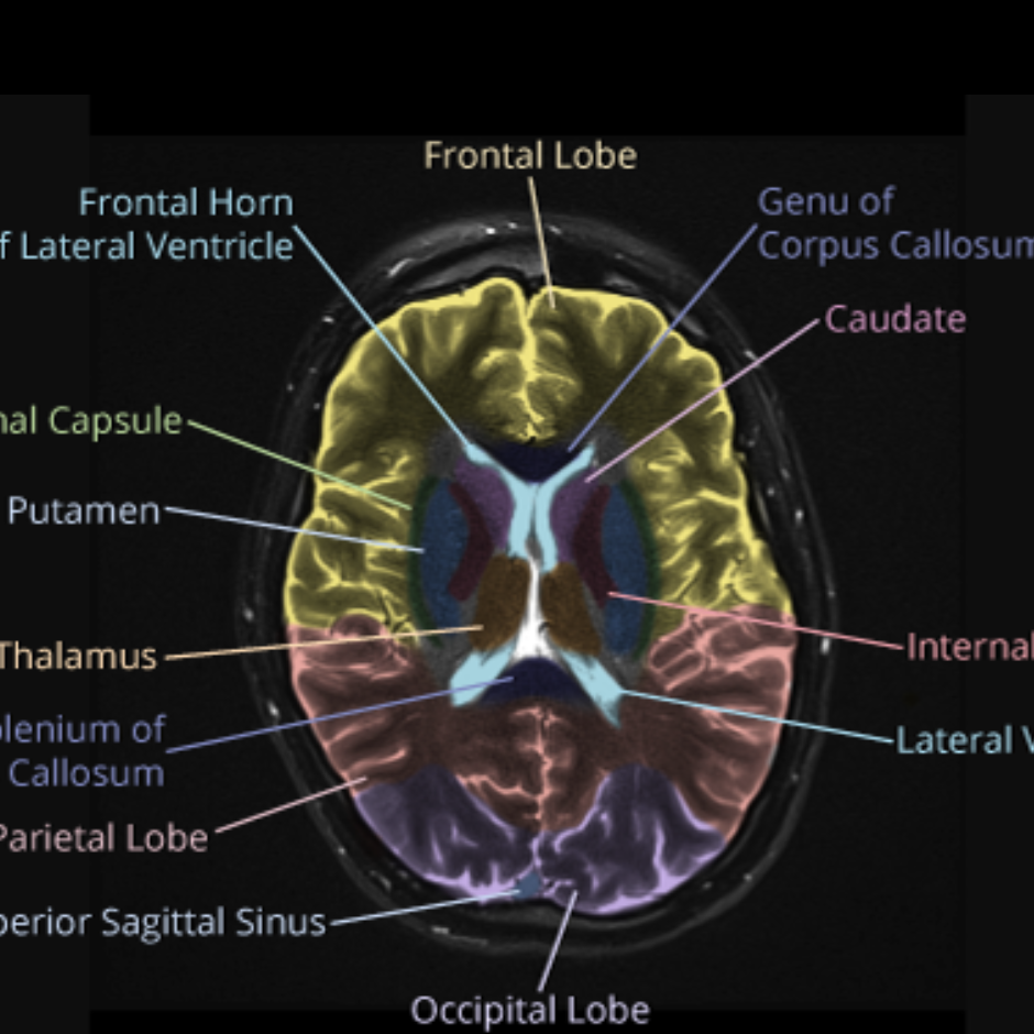
CT Brain AnatomyGrey matter structures. The cortex, insula, basal ganglia and thalamus are the important grey matter structures. Important grey matter structures visible on CT images of the brain include the cortex, insula, basal ganglia, and thalamus.

Brain lobes annotated MRI Image
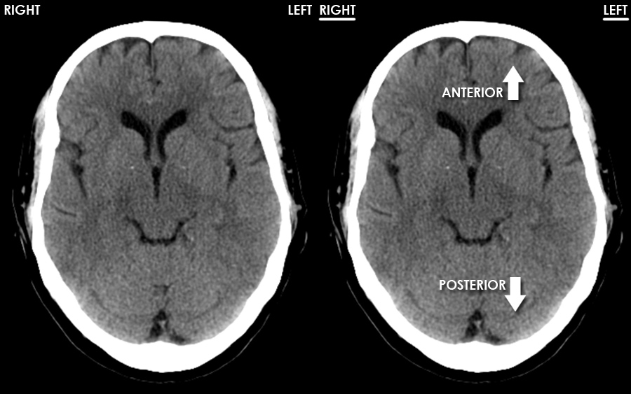
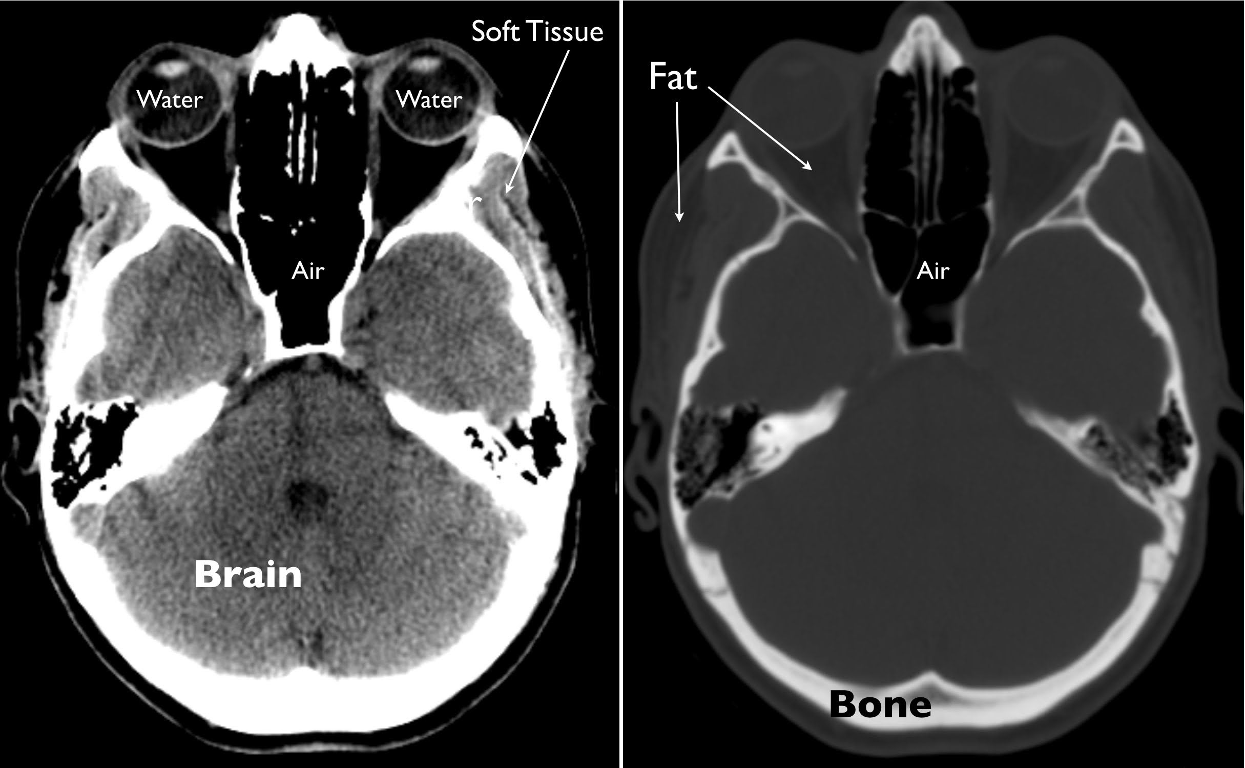
CT images of the brain are conventionally viewed from below, as if looking up into the top of the head. This means that the right side of the brain is on the.
CT Brain Anatomy Tutorial introduction
ANATOMICAL PARTS Ala of nose Alveolar process Ambient cistern Angular gyrus Anterior arch of atlas Anterior cerebral artery: Postcommunicating part; A2 segment Anterior cerebral artery: Precommunicating part; A1 segment Anterior chamber Anterior clinoid process Anterior commissure Anterior communicating artery
Normal anatomy of the brain on CT and MRI with a few normal variants Practical Neurology
The brain is surrounded by cerebrospinal fluid (CSF) within the sulci, fissures and basal cisterns.CSF is also found centrally within the ventricles.The sulci, fissures, basal cisterns and ventricles together form the 'CSF spaces', also known as the 'extra-axial spaces'. CSF is of lower density than the grey or white matter of the brain, and therefore appears darker on CT images.

Ct Scan Brain Anatomy Anatomy Of Head Ct Scan Normal The Brain On Ct And Mri / Frontal
The video shows the basic CT anatomy of the brain.For each slice we have highlighted. This video is a part of basic radiologic head CT SCAN anatomy series. The video shows the basic CT anatomy.
Exploring the Brain How Are Brain Images Made with CT? UCSF Radiology
ct Normal CT head with annotated and original images. Case Discussion Annotated teaching CT head in standard and bone windows. 62 public playlists include this case (advertising)

Brain and face CT interactive anatomy atlas eAnatomy
CT images of the brain are conventionally viewed from below, as if looking up into the top of the head. This means that the right side of the brain is on the left side of the viewer. The anterior part of the head is at the top of the image. CT brain - image orientation Hover on/off image to show/hide findings Click image to align with top of page

The Radiology Assistant Brain Anatomy
CT head (sometimes termed CT brain ), refers to a computed tomography examination of the brain and surrounding cranial structures. It is most commonly performed as a non-contrast study, but the addition of a contrast-enhanced phase is performed for some indications. This article covers non-contrast and delayed post-contrast imaging.
CT Scan Tips & Protocols CT BRAIN ANATOMY
CT Brain ct Axial non-contrast C+ delayed C+ delayed C+ delayed Brainstem and cerebellum without evidence of focal lesions. Lateral ventricles of normal volume. Third and fourth ventricles in midline. Basal subarachnoid cisterns normal configuration. Focal abnormalities are not observed in the brain parenchyma.